Digital pathology and deep learning, project bases to identify breast cancer
Santa María Tonantzintla, Puebla, October 26, 2023.- Optimizing breast cancer diagnosis methods based on the analysis of the tumor microenvironment through automated analysis of digital images, is one of the objectives of a project of the National Institute of Astrophysics, Optics and Electronics (INAOE), research center of the National Council of Humanities, Sciences and Technologies (Conahcyt).
This is an interdisciplinary project in which researchers and students of Computer Sciences and Biomedical Sciences and Technologies from INAOE, and doctors and specialists from the Jalisciense Institute of Cancerology and the Center for Industrial Technical Education (CETI), of Jalisco, work.
The project arose from collaboration with researchers from Jalisco, Dr. José Cruz Ramos and Dr. Gabriela López Armas. Two years ago, a first master's thesis in Biomedical Sciences and Technologies was developed at INAOE. Currently Héctor Zepeda Reyes and Miguel Loria Romero, doctoral students at INAOE, are working on the topic.
Dr. Hayde Peregrina Barreto, INAOE researcher and project leader, reports that she is working with public databases of digital samples of cancer histopathological sections in which there is suspicion of cancer.
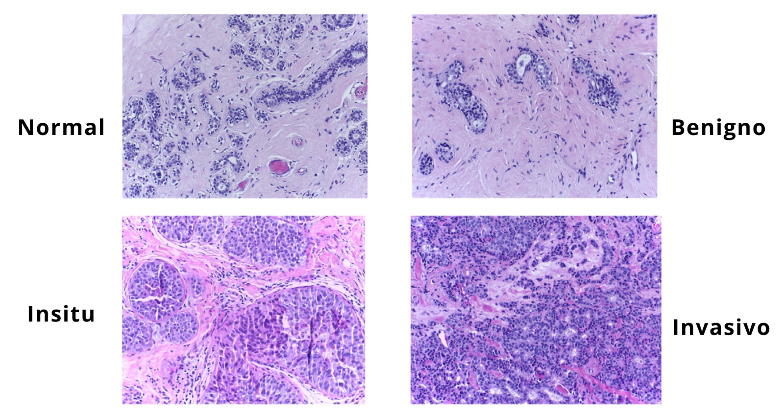
“From the biopsy, cuts are made to analyze small fragments of the tissue and see how present the malignancy is or confirm it. In this tissue, the structures of the cells and other specific structures of breast cancer such as the tubules, the cells that are already doing mitosis and the nuclei are studied,” she comments.
The project seeks to automate this analysis and quantify the variation of the structures, a procedure that the pathologist traditionally does by observing the microscope and based on experience.
“Many studies have concluded that there may be significant discrepancy between pathologists. To have a more precise reference of what is being evaluated, digital methodologies for digital image analysis have begun to be developed to support this process in which the pathologist observes a certain number of fields or spaces on the sample and On each one of them he makes counts, determines the shape, and analyzes those structures. It is a time-consuming task. If we can automate at least part of the task, we would help the pathologist by avoiding fatigue and reducing time and costs.”
Héctor Zepeda Reyes, a doctoral student in Biomedical Sciences and Technologies at INAOE, has become involved in the project thanks to his training as a molecular biologist. “Working on breast cancer always caught my attention. As I already had some work during my master's degree with image processing, I proposed to Dr. Hayde an idea that she already had, and the doctor already had the background of her master's classmate. My project is focused above all on stromal analysis. The stroma behaves like the cellular environment. Although when making the diagnosis the pathologist focuses on structures such as cells, mitosis and lumina, there is a new approach that tries to evaluate how the cellular environment behaves and what its structural patterns are, because it is closely related with the development and malignancy of cancer. Evaluating this with image processing and artificial intelligence tools will help us have better precision when diagnosing and offer better tools to specialists,” he says.
In 2024, Héctor Zepeda will spend a stay at the Jalisciense Institute of Cancerology to begin labeling all the samples and working, together with the pathologist, on a database of Mexican women.
Dr. Hayde Peregrina notifies that with another of her doctoral students, Miguel Loria Romero, and in collaboration with a researcher from the University of Alcalá, Spain, they are working on the development of a tool focused on the education or specialized training of pathologists. . She also points out that these types of projects depend on strong collaboration with her medical peers.
"Given that there may be discrepancies between pathologists, we want to make a system that evaluates what they observe and then through a deep learning model we can tell if it failed or if it was missing any element, which will allow us to have a metric of How your training is improving. Deep learning allows us to gather information, which already has a label, and feed a model that can learn with the help of the information given by the pathologist and that allows us to refine the results, being able to be more reliable,” she underlines.
Finally, Héctor Zepeda indicates that they already have interesting results and that the range of ideas and analysis is expanding: “From the biological approach, this combination between the computational and the biological part will define the future of the biomedical area.”
Luis Enrique Erro # 1, Tonantzintla, Puebla, México, Código Postal 72840, Tel: (222) 266.31.00, difusion@inaoep.mx
This work is licensed under a Creative Commons Attribution-NonCommercial-NoDerivs 2.5 Mexico License.
